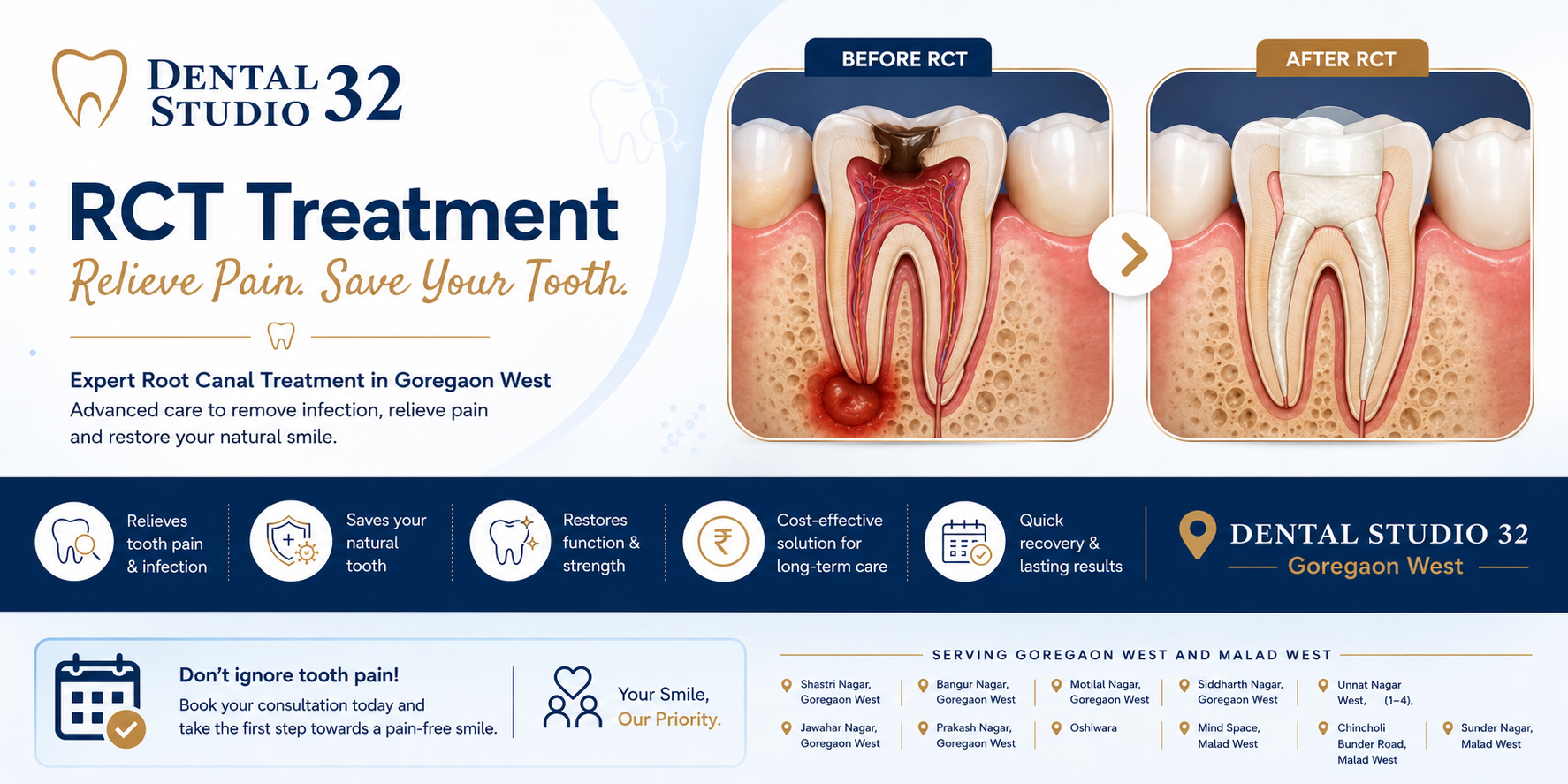
The Situation — A Molar in Crisis
Our patient was a 34-year-old working professional from Oshiwara who had been managing intermittent toothache in her lower right jaw for the better part of eight months. The pain was inconsistent — sometimes sharp and sudden, sometimes a dull throb that lasted for hours, occasionally absent entirely. That inconsistency had made it easy to delay.
By the time she visited the first clinic, a localised infection had spread into the root canals of her lower right first molar. The X-ray showed extensive decay reaching the pulp, widening of the periodontal ligament space, and a developing periapical abscess — a pocket of infection at the root tip. Both clinics she visited advised extraction as the straightforward path forward and made recommendations for replacement options, including an implant.
She found us through a Google search — specifically searching for a dentist in Goregaon West who could give her a second opinion. She came in not hoping for a different answer. She came in wanting to understand whether there was one.
"I wasn't expecting you to tell me something different. I just wanted to understand why my tooth couldn't be saved before I agreed to lose it."Our patient, 34 — Oshiwara, Goregaon West
The Clinical Assessment — What We Found
Dr. Stutee reviewed the existing X-rays, took fresh periapical and bitewing radiographs, and performed a detailed clinical examination including percussion, palpation, and thermal sensitivity testing. The diagnosis confirmed severe irreversible pulpitis with periapical pathology — in plain terms, the pulp tissue inside the tooth was non-vital and the infection had begun to affect the bone at the root tips.
However, the key clinical question was not whether the tooth was infected — it clearly was. The question was whether adequate tooth structure remained after decay removal to support a successful root canal and long-term restoration. The answer, in Dr. Stutee's assessment, was yes — with a clear margin.
💡 A note from Dr. Stutee: Root canal treatment has a long-standing and largely undeserved reputation for being the worst-case scenario. In clinical practice, it is often the exact opposite — the option that preserves a natural tooth, avoids the cost and complexity of an implant, and resolves infection with precision. When a tooth can be saved, saving it is always the preferred path. The decision to extract should be reached only when the tooth genuinely cannot be restored to function.
Why This Case Required Careful Planning
Root canal treatment on a lower first molar is among the more demanding endodontic procedures in general practice. The tooth has multiple canals — typically three or four — that must each be located, instrumented to their full length, thoroughly irrigated, and hermetically sealed. Any canal that is missed, inadequately cleaned, or poorly obturated becomes a site for reinfection.
Clinical Complexities
Three canals with moderate curvature requiring precise rotary instrumentation. Active periapical infection requiring resolution before obturation. Deep mesial decay demanding careful access cavity design to preserve structural integrity throughout.
Factors in Our Favour
Intact alveolar bone with no vertical loss. Localised infection with no systemic involvement. Sufficient coronal tooth structure for full crown restoration. Patient motivated and committed to completing the full treatment sequence.
The infection also meant that treatment had to be staged. Beginning obturation — the sealing phase — before the infection had fully resolved would risk trapping bacteria and causing a flare-up or treatment failure. The sequencing of sessions was therefore as important as the technical execution within each one.
The Procedure — Session by Session
Root canal treatment unfolds across carefully sequenced appointments. For a case with active periapical infection, rushing through to completion in a single extended session is not best practice — it is a shortcut that compromises the result. Our patient's treatment was completed across three appointments, each serving a distinct clinical purpose.
Diagnosis, CBCT Imaging, and Access Opening
Comprehensive clinical and radiographic examination including cone beam CT to map all root canals and assess bone levels precisely. Access cavity preparation under rubber dam isolation — a sterile barrier that prevents oral bacteria from contaminating the working field during the procedure. Working lengths established for all three canals using an electronic apex locator and confirmed radiographically. Initial irrigation with sodium hypochlorite to begin disinfection. Intracanal medicament placed to address the active infection between sessions.
Rotary Canal Shaping and Thorough Irrigation Protocol
Rotary nickel-titanium files used to shape each canal to its full working length while preserving natural root curvature — critical for avoiding procedural errors such as ledging or transportation. Multi-irrigant protocol using sodium hypochlorite, EDTA, and final rinse with chlorhexidine to ensure comprehensive debridement of organic and inorganic debris from within the canals. Clinical review confirmed symptom resolution and absence of sinus tract. Periapical X-ray showed reduction in abscess shadow. Second intracanal dressing placed for additional biological healing time.
Canal Sealing and Post-Endodontic Crown Preparation
Canals dried and obturated with gutta-percha and bioceramic sealer using warm vertical compaction — a technique that achieves a dense, void-free three-dimensional fill within the root system. Radiographic confirmation of complete obturation taken immediately post-procedure. Definitive coronal seal placed. Crown preparation also completed at the same appointment — a deliberate clinical decision, as delaying the permanent crown following obturation significantly increases the risk of reinfection through coronal leakage. A full porcelain-fused-to-metal crown was fabricated and cemented within two weeks.
4 weeks total
including setup
to final crown cementation
complications
The Result — A Tooth That Works, a Patient Who Kept It
At the three-month follow-up appointment, periapical X-rays confirmed complete resolution of the abscess and healthy periapical bone. The crowned tooth was functioning normally with no sensitivity, no swelling, and no residual discomfort. The patient reported being able to chew on the right side again for the first time in over a year.
The clinical outcome is straightforward to summarise: the tooth is saved, the infection is gone, and the crown will protect that tooth for many years. But the broader point of this case is what it illustrates about the diagnostic process — specifically, that an extraction recommendation reflects a clinical judgment, not an unavoidable fact. That judgment can be accurate. It can also, in cases like this one, be worth a second look.
"At the follow-up, she showed me the X-ray and pointed to where the abscess had been. Then she pointed to where it wasn't anymore. That was a good appointment."Dr. Stutee Nikhil Shukla — Dental Studio 32, Goregaon West
Common Misconceptions About Root Canal Treatment
Root canal treatment is one of the most misunderstood dental procedures. Patients frequently delay or decline it based on information that is decades out of date. These are the most common ones we encounter — and the clinical reality behind each.
Root canal treatment is extremely painful. This belief originates from pre-anaesthetic dentistry. With modern local anaesthesia, the procedure itself is comfortable — often more comfortable than the toothache that preceded it. The discomfort associated with RCT is overwhelmingly the infection, not the treatment.
The treatment relieves the pain caused by infection. The goal of root canal treatment is to remove the infected tissue, eliminate bacteria from within the root system, and seal the canal. Patients routinely report significant relief within 24–48 hours of the first appointment.
Extracting a tooth is simpler and equally effective. Extraction removes the immediate problem but creates a secondary one: a gap in the arch. An untreated gap leads to drifting of adjacent teeth, bone resorption at the extraction site, bite changes, and — eventually — the need for more complex restorative work. Natural teeth, when they can be saved, should be.
A saved tooth with a crown outperforms most replacements in the long run. A successfully treated and crowned natural tooth can last as long as the patient looks after it. It preserves the bone, maintains the bite, and requires no special maintenance beyond normal oral hygiene.
Root canal treated teeth are fragile and fail quickly. The concern is understandable — the tooth is devitalised, meaning it no longer has a blood supply. However, a correctly obturated root canal followed by a well-fitted crown provides a durable, fully functional restoration with high long-term success rates when patients maintain regular reviews.
Success rates for root canal treatment in correctly selected cases are high. Clinical studies consistently report success rates above 90% at five-year follow-up for initial root canal treatment in suitable cases. The most common cause of failure is delayed or absent coronal restoration — which is why we place the crown promptly.
Signs You May Need Root Canal Treatment
Root canal treatment is indicated when the pulp — the inner tissue of the tooth containing nerves and blood vessels — is infected or dying. The symptoms can vary, and some patients have very little pain even with significant infection. If you recognise any of the following, a prompt examination at our dental clinic in Goregaon West is advisable.
⚠️ Do not ignore these symptoms. Dental infections do not resolve on their own. Delayed treatment increases the risk of abscess spread, bone loss, systemic involvement, and — ultimately — losing a tooth that could have been saved with timely care.